
Tubular breasts are almost always described as small breasts. That description is the reason the wrong operation gets planned.
Many patients with a tubular pattern have adequate or even generous breast tissue. The problem is that the tissue is distributed inside a constricted framework that prevents the breast from assuming a natural, rounded contour. It is not a size problem. It is an architecture problem — and that single distinction determines why standard augmentation alone frequently fails here, and why the plan has to address the structural pattern rather than simply adding volume.
What the tubular pattern actually is
Tubular breast deformity — also called constricted breast — is a developmental variation in breast shape. The breast base is narrow, the lower pole is underdeveloped or tethered, and breast tissue can herniate into an enlarged areola. Patients usually describe the result as elongated, tubular, or disproportionately projected relative to how little of the chest wall the breast actually occupies.
The degree varies along a spectrum. Some patients have mild base constriction with modest lower-pole deficiency. Others have severe tubularity with significant areolar herniation and marked asymmetry between the two sides. Grading that spectrum accurately is the first clinical task, because the plan changes considerably along it.
A narrow footprint holding a normal amount of tissue
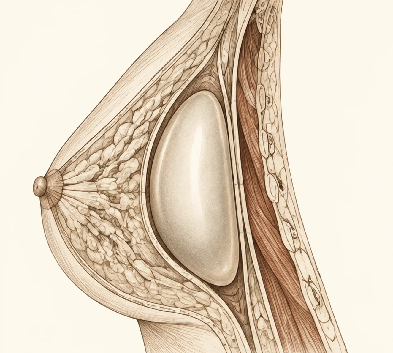
The breast base is the footprint where the breast attaches to the chest wall. In a tubular breast that footprint is narrower than it should be for the volume of tissue present, and a ring of tight tissue at the breast’s periphery prevents the lower pole from expanding during development. Instead of filling out into a rounded lower curve, the tissue is pushed upward and forward — producing a narrow, projecting shape with an empty or deficient lower pole. In many patients the tissue then herniates through the areola, the path of least resistance, creating an enlarged, puffy or dome-shaped areolar complex.
Why the operation is a reshaping procedure, not an augmentation
Tubular breast correction is structural reshaping surgery designed to address the constricted base, redistribute tissue into a more natural contour, manage areolar prominence where it is present, and restore proportion between the breast and the chest wall.
It is not a single technique. It is a category of surgical strategies selected and combined according to the anatomy in front of me. The plan has to address the mechanism of the deformity rather than its appearance, because the visible shape is a consequence of the underlying architectural constraint. Treating the surface without addressing the foundation produces results that look incomplete or unnatural.
Releasing the constricted base comes first
In most cases the first element of correction is releasing the tight ring of tissue that prevents lower-pole expansion, so that the breast can take up a wider and more natural footprint. Until that is done, volume added to the breast — whether by implant or by fat grafting — simply inflates the existing tubular shape instead of correcting it.
Once the constriction is released, the native tissue is free to redistribute and the overall shape can emerge coherently. This is the sequence that decides the result, and it is the sequence most often skipped.
The most common planning error is treating a constricted breast as an underfilled one
An implant placed behind a constricted base can produce a “double bubble” — the implant creates one contour while the constricted native tissue creates another, visible as a step-off or shelf across the lower breast. The tissue was never simply small. It was imprisoned within a constrictive architecture, and adding volume to an unreleased framework makes the underlying pattern more obvious rather than less.
Volume support is a decision, not a default
Volume is not always necessary, and when it is, the method should be chosen by mechanism rather than by habit.
Some patients have adequate tissue volume that only needs redistributing into a corrected base — once the constriction is released and the lower pole expands, the existing tissue fills the new shape acceptably. Others have genuine volume deficiency in addition to the architectural problem, and benefit from augmentation with an implant or with autologous fat transfer. The choice depends on how much volume is needed, the quality and elasticity of the overlying tissue, body habitus, and the trade-off profile each method carries.
Three volume strategies after the base has been released
| Feature | Release and redistribution alone | Release plus implant | Release plus fat grafting |
|---|---|---|---|
| When it is chosen | Tissue volume is adequate and only needs to fill a corrected base | There is genuine volume deficiency in addition to the constriction | Additional volume is needed, or transitions require softening and subtle contour refinement |
| What it delivers | A wider footprint and a more naturally distributed contour from existing tissue | Predictable, defined volume | Tissue-integrated augmentation that blends into the native breast |
| Principal trade-off | The achievable volume is limited to what the patient already has | Device-related considerations: pocket behaviour, long-horizon durability, and the interaction between the implant and the constriction-corrected tissue | Fat survival is variable, so the outcome is less precisely predictable |
| Main limitation | Insufficient where volume deficiency is a real part of the problem | Cannot compensate for an inadequately released base | Achievable volume per session is limited |
Neither augmentation method is universally superior. The correct choice is the one that matches the anatomical need — and in a meaningful number of patients, the correct choice is no added volume at all.
The areola is usually part of the deformity, not a separate request
Areolar herniation and enlargement are hallmark features of the tubular pattern. When breast tissue has been pushed through the areola by the constricted base, the areola appears puffy, dome-shaped, or disproportionately large relative to the breast mound.
Reducing areolar diameter and reshaping the areolar complex restores proportion between the nipple–areola unit and the surrounding breast. This introduces periareolar scars — scars around the areolar border — which generally heal well but are subject to the same biological variability that affects all scars. In some patients additional envelope management is needed, introducing scars in patterns similar to those used in breast lift surgery.
Meaningful shape correction here requires accepting some visible incision work
The scar trade-off has to be stated plainly rather than softened. A patient who needs a significant shape change but is unwilling to accept any scarring faces a genuine limitation, and that limitation may reduce the achievable result. This is not a reason to decline the operation; it is a reason to decide with the trade-off in view rather than discover it afterwards.
Asymmetry is the rule, not the exception
The two breasts frequently differ in degree of constriction, lower-pole development, areolar size and overall volume. Correcting both sides to look identical is a goal the plan pursues but that biology does not guarantee.
Differential healing, differential tissue elasticity and the inherent asymmetry of the starting anatomy all contribute to residual differences that may persist. Symmetry is a goal, not a promise. A patient who requires absolute mirror-image symmetry as a condition of satisfaction will find this operation a frustrating experience, and I would rather say that at the consultation than at the follow-up.
I grade the constriction before I discuss volume
My planning order in this operation does not change: identify the tubular pattern, grade the base constriction and lower-pole deficiency, assess the areolar component and the asymmetry between sides, and only then ask whether volume is needed and by which method. Adding volume is the easiest part of this operation to discuss and the least likely part to determine the result. The best outcomes come from accurate diagnosis of the constriction pattern and conservative reshaping that respects tissue limits — not from aggressive correction pursuing a template ideal.
Recovery is a sequence, not a single date.
- Early phaseSwelling exaggerates the correction
Swelling is expected and can temporarily make the breast look fuller or more projected than the final result. This is the least reliable period for judging shape.
- Lower-pole expansionReleased tissue settles into a new architecture
As oedema resolves, the lower pole — released from its constricted position — needs time to expand and soften into a natural curve. As I tell patients: the breast at six weeks may still look quite different from the breast at six months.
- Scar maturationThe incision lines mature on their own timeline
Scars typically progress from an initially red or raised appearance toward a flatter, lighter line over many months, independently of how the shape itself is settling.
I avoid fixed timelines because healing depends on tissue quality and individual tissue behaviour. Patients who understand this staged evolution assess their result at appropriate intervals instead of drawing conclusions from an incomplete healing process.
What this operation cannot deliver
It cannot guarantee a specific template breast shape. Tissue quality, skin elasticity and healing biology set ceilings that differ from patient to patient.
It cannot produce a scarless result when the correction requires envelope management or areolar reduction. It cannot always be completed in a single operation — severe tubular deformities with marked asymmetry may benefit from staged correction, where the primary surgery establishes the structural framework and a secondary refinement addresses residual shape or symmetry once the tissues have fully settled. And it cannot stop the breast from continuing to change: ageing, weight fluctuation, hormonal changes and gravity all continue to influence breast shape after surgery, exactly as they do in unoperated breasts.
Why the first operation should establish the framework
Revision in a previously corrected tubular breast operates under more constrained conditions. Scar planes from the initial release and from any prior augmentation or lift alter the tissue environment, and blood supply patterns may be less robust. The tissue can also exhibit structural memory — a tendency to contract back toward its original constricted configuration despite surgical release.
Revision goals therefore have to be more specific and more conservative than primary goals, and the decision to operate again must weigh meaningful improvement against the diminishing returns and increasing complexity that characterise secondary breast surgery.
What should be weighed in the decision?
This operation reshapes constricted tissue, which means the trade-offs are structural rather than incidental. The items below are the ones that most often decide whether a patient should proceed, stage the correction, or wait.
- Trade-off: meaningful shape correction typically requires visible incision work. Areolar reduction introduces periareolar scars, and envelope management introduces scars in patterns similar to breast lift surgery.
- Trade-off: scars generally heal well but remain subject to individual biological variability.
- Trade-off: an implant provides predictable, defined volume in exchange for device-related considerations — pocket behaviour, long-horizon durability, and the interaction between the implant and the constriction-corrected tissue.
- Trade-off: fat grafting integrates into the tissue and softens transitions, but fat survival is variable and the achievable volume per session is limited.
- Trade-off: staging a severe deformity means two operations rather than one, in exchange for a more reliable final shape.
- Limitation: asymmetry is the rule in tubular breasts. Residual differences may persist and are within the expected range.
- Limitation: individual tissue behaviour — how skin contracts, how released tissue redistributes, how scars mature, how implants or grafted fat settle — introduces variability no technique fully controls.
- Limitation: a specific template breast shape cannot be guaranteed; tissue quality and skin elasticity set the ceiling.
- Limitation: a scarless result is not achievable when envelope management or areolar reduction is required.
- Limitation: under-correction is a real risk, particularly when the base constriction is not adequately released.
- Limitation: contour irregularity is possible after reshaping constricted tissue.
- Limitation: a “double bubble” step-off can occur when volume is added behind a constricted base.
- Limitation: severe deformities may not be correctable in a single operation.
- Limitation: revision is constrained by altered scar planes, potentially less robust blood supply, and the tissue’s tendency to contract back toward its constricted configuration.
- Limitation: the breast continues to change with ageing, weight fluctuation, hormonal change and gravity after surgery.
- Alternative: where tissue volume is adequate, release and redistribution without any added volume is the appropriate plan.
- Alternative: where the deformity is severe and asymmetric, staged correction is preferable to attempting everything at once.
- Alternative: where absolute mirror-image symmetry or a scarless outcome is a condition of satisfaction, this operation is not the right decision at this time.
How to think about the decision
Correction is properly indicated when the tubular pattern is clearly identified, when the plan addresses the architectural mechanism rather than the surface appearance, and when the scar trade-offs and biological variability inherent in reshaping constricted tissue are understood and accepted.
Under those conditions the improvement can be significant. A narrow, elongated breast can be converted into a more rounded, naturally distributed contour. Proportion between the areola and the breast mound can be restored. Bra fit and clothing drape improve, as does the patient’s relationship with a body feature that has often been a source of distress since adolescence.
What makes that outcome reliable is not the intensity of the correction. It is accurate diagnosis of the constriction pattern, conservative reshaping that respects tissue limits, and the recognition that coherent breast architecture — a shape that looks natural at rest and in motion — is a more durable goal than photographic perfection.
How do I know if I have tubular breasts?
Common features include a narrow breast base, an underfilled or tethered lower pole, enlarged or puffy areolae, and herniation of breast tissue into the areolar complex. The degree varies along a spectrum — from mild base constriction to severe tubularity with marked asymmetry. An in-person assessment determines the grade and guides whether correction is indicated and what approach fits your anatomy.
Is this a size problem or a shape problem?
A shape problem. Many patients with tubular breasts have adequate or even generous breast tissue, but it is distributed within a constricted framework that prevents a rounded contour. This distinction matters practically, because it is the reason augmentation alone frequently fails to produce a satisfying result here.
Why is standard breast augmentation not enough?
Because volume added to an unreleased framework inflates the existing tubular shape rather than correcting it. The tight ring of tissue restraining the lower pole has to be released first so the breast can take up a wider, more natural footprint. Adding volume behind a constricted base can also create a “double bubble” — a visible step-off across the lower breast where the implant contour and the constricted native tissue contour disagree.
Do I need implants?
Not always. Some patients have adequate tissue volume that simply needs to be redistributed once the constriction is released. Others have genuine volume deficiency and benefit from implants or fat grafting. The choice depends on the degree of correction needed, tissue quality, and the specific trade-off profile of each method — not on a default assumption that augmentation is required.
Implant or fat grafting — which is better?
Neither is universally superior. Implants provide predictable, defined volume but introduce device-related considerations such as pocket behaviour and long-horizon durability. Fat grafting integrates into the tissue and is particularly useful for softening transitions and refining contour, but fat survival is variable and the achievable volume per session is limited. The correct choice is the one that matches the anatomical need.
Will the areola be reduced?
Often, yes — if areolar herniation or enlargement is part of the deformity. Reducing the diameter and reshaping the areolar complex restores proportion between the nipple–areola unit and the breast mound. This introduces periareolar scars, which generally heal well but are subject to individual tissue behaviour.
Can it be done in one operation?
Often, but not always. Severe tubular deformities with marked asymmetry may benefit from staged correction, where the primary surgery establishes the structural framework and a secondary refinement addresses residual shape or symmetry once the tissues have fully settled. Staging is a planning decision made in the interest of the final shape, not a sign that something went wrong.
My two breasts are quite different. Can they be matched?
Asymmetry is the rule rather than the exception in tubular breasts, and the two sides frequently differ in constriction, lower-pole development, areolar size and volume. The plan actively addresses this, but differential healing and differential tissue elasticity mean residual differences may persist. Symmetry is a goal, not a promise.
How variable is recovery?
Recovery is staged. Swelling can temporarily exaggerate the correction, and the lower pole needs time to expand and soften into a natural curve after constriction release. Early shape is not final shape — the breast at six weeks may look quite different from the breast at six months. I avoid fixed timelines because healing depends on tissue quality and individual tissue behaviour.
What are the main risks?
Risks include asymmetry, scarring issues, under-correction, contour irregularity, and the possibility that staged revision may be needed for severe deformities. A common planning error is treating a tubular breast as simply small rather than structurally constricted — which can produce a “double bubble” or an inflated tubular shape instead of a natural contour.
Can it be revised later if I am not satisfied?
Revision is possible but operates under more constrained conditions. Scar planes from the initial release and any prior augmentation or lift alter the tissue environment, blood supply patterns may be less robust, and the tissue can contract back toward its original constricted configuration. Revision goals therefore need to be more specific and more conservative than primary goals.
What should I realistically expect?
You should expect improved breast shape and proportion — a more rounded, naturally distributed contour with a balanced areola — not perfect symmetry or a scarless outcome. Asymmetry is the rule in tubular breasts, and while it is actively addressed, residual differences are within the expected range. A thorough assessment clarifies what can be achieved and where the biological limits are.